My Store
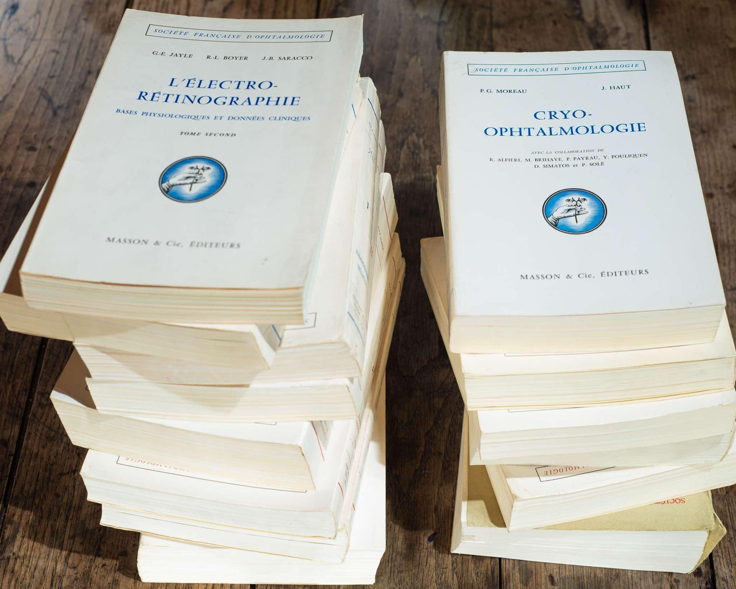
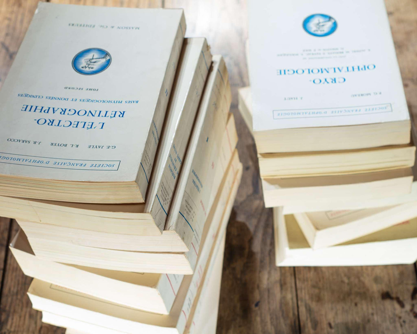
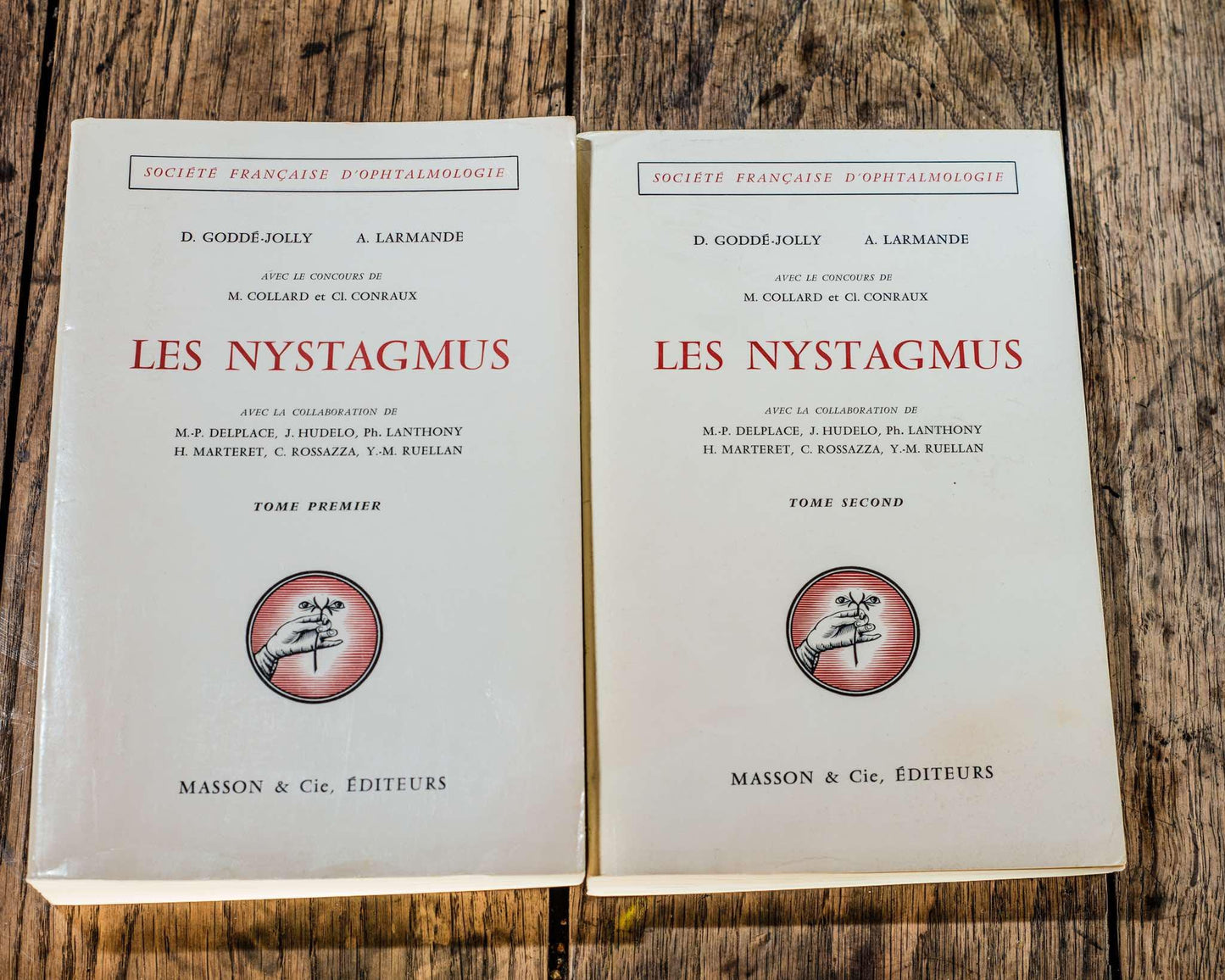
The set of 13 medical books, ophtalmology 1964-1973, Masson et cie, èditeurs 120, Medicine, Textbook for Doctors and Students Study Book
The set of 13 medical books, ophtalmology 1964-1973, Masson et cie, èditeurs 120, Medicine, Textbook for Doctors and Students Study Book
Regular price
€700,00 EUR
Regular price
Sale price
€700,00 EUR
Unit price
per
Taxes included.
Couldn't load pickup availability
The set of 13 medical books includes:
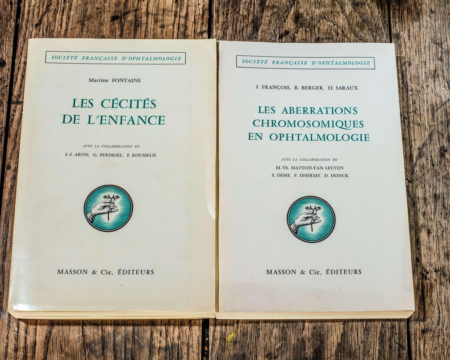
CHROMOSOMAL ABERRATIONS IN OPHTHALMOLOGY
AUTHORS:
J. FRANCOIS, R. BERGER, H. SARAUX, M.TH. MATTON-VAN LEUVEN, S. DEBIE, P. DHERMY, D. DONCK
PARTS:
Cytogenetic elements
Autosome abnormalities
Sex chromosome abnormalities
Triploidies
Report presented to the French Society of Ophthalmology on May 9, 1972
Masson & CIE, èditeurs 120, boulevard Saint-Germain, Paris (vie)
FOREWORD
First of all, we would like to express our gratitude to the Committee and Members of the French Society of Ophthalmology for the great honor they have done us and for the kind esteem they have shown us, by entrusting us with the writing of the Report on Chromosomal Aberrations in Ophthalmology.
We would particularly like to thank Professor DUBOIS-POULSEN, who had this subject accepted by the General Assembly of 1967 and appointed us as rapporteurs. Our gratitude also goes to Professor DESVIGNES and the current management of the Society, who have never ceased to show us the most friendly kindness and the most cordial concern. We hope not to disappoint too much the trust that their friendship has placed in us.
We owe special gratitude to all those who have very kindly communicated their work, their material, their iconography or their clinical and histopathological observations: Pr ARDOUIN (Rennes), Pr AUSSANNAIRE (Paris), Pr CH. BACH (Paris), Dr M. L. BARR (London, Canada), Mrs Pr M. BRIHAYE-VAN GEERTRUYDEN (Brussels), D' J. BRODY (Melbourne), Dr B. CAGIANUT (Zurich), DE A. DE LA CHAPELLE (Helsinki), Pr P. DANIS (Brussels), Pt C. DUPUIS (Lille), Dr D. GODDE-JOLY (Paris), Pr J. DE GROUCHY (Paris), D'. K. H. GUSTAVSON (Upsala), Dr. K. HEIMANN (Cologne), Pr. J. R. MILLER (Vancouver), Pr P. MozZI- CONACCI (Paris), Dr E. ORYE (Ghent), Dr C. E. PARKER (Los Angeles), P' A. PRADER (Zurich), Dr N. RICCI (Ferrara), Pr H. ROELS (Ghent), Dr A. TARKKANEN (Helsinki), Dr A. TUMBA (Leuven), P H. VAN DEN BERGHE (Leuven), Dr M. YANOFF (Washington).
The valuable collaboration of all these eminent researchers has greatly facilitated our task and enriched our documentation
For two of the rapporteurs, the bulk of this work could only be achieved thanks to the assistance and friendship of Father JEROME LEJEUNE, it is his observations and his iconography that are the basis of a whole part of this report. We would like to thank him and to associate his entire team with these thanks, we owe, among others, a lot to doctors M. O. RÉTHORÉ, B. DUTRILLAUN, M. PRIEUR, but this list is not exhaustive.
Thanks also to all our immediate collaborators, who all, in one way or another, contributed with as much generosity as knowledge to the development and writing of this report, thanks to them, it has become a team effort. We are deeply grateful to them.
Although by force of circumstances the writing of each chapter was individual, the most complete agreement has never ceased to exist between the rapporteurs. Each of us contributed to the others his documentation, advice and criticism. Each chapter was reviewed by the other two contributors, so that the entire report is truly a team effort, representing a common understanding of the issues raised.
We apologize in advance for any imperfections in our report and count on the indulgence of our readers. We hope, however, that it will not be a blot on the illustrious work that has contributed for many years to the worldwide influence of the most famous of the National Societies.
INTRODUCTION
It seemed essential to us to give at the beginning of this report on chromosomal aberrations in ophthalmology some fundamental notions of cytogenetics in order to make ocular malformations due to chromosomal anomalies more understandable. It is, in fact, not useless to recall the examination techniques, the morphology of dermatoglyphs, the characteristics of the normal human karyotype, the different types of chromosomal aberrations, their frequency and their etiology, if we wish to deepen the ocular pathology of chromosomal origin.
We could have studied the ocular manifestations starting from each symptom or from each anatomical part of the eyeball and its adnexa. Our attempt at synthesis would have been very uninformative and all the more confusing, since the severity and frequency of ocular anomalies are very variable according to the chromosomal group modified by excess or by defect. This is why we preferred to study successively the anomalies of the different groups of autosomes and of the group of gonosomes by detailing for each of them the ophthalmological manifestations and by insisting on their relative frequency and importance.
The amount of pages: 194
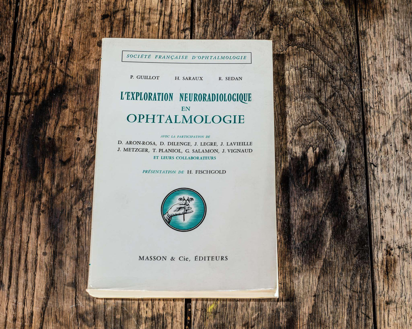
NEURORADIOLOGY EXPLORATION IN OPHTHALMOLOGY
AUTHORS:
D. ARON-ROSA, J. METZGER, D. DILENGE, T. PLANIOL, J. LEGRE, G. SALAMON, J. LAVIEILLE, J. VIGNAUD
PARTS:
1. Neuroradiology anatomy of the optic pathways
2. Exploration methods
3. Indications and choice of techniques
4. Accidents
Report presented to the French Society of Ophthalmology on May 10, 1966
Masson & CIE, èditeurs 120, boulevard Saint-Germain, Paris (vi
INTRODUCTION
THE COMMITTEE OF THE FRENCH SOCIETY OF OPHTHALMOLOGY asked us, 5 years ago, to study the "ophthalmological complications of neurosurgical exploration procedures".
It quickly became apparent to us that our work, thus conceived, would call for at least two criticisms:
criticism of its negative character, the conclusion of such a study risking arousing in the ophthalmologist an unfounded fear of neuroradiology, pushing him to restrict its indications, thus going against all that is progress in modern medicine;
criticism of its character too "historical", neurosurgical exploration methods having been considerably improved in recent years, most of the complications described in the literature relate, in fact, to examinations carried out with the means of the past.
This rapid evolution of techniques has also led us to note that there was no comprehensive work on neuroradiological explorations in the ophthalmological literature.
It is in an attempt to fill this gap that, guided by the inexhaustible benevolence of Professor H. FISCHGOLD, we have produced this report, which is intended to be a provisional update of what neuroradiology can bring to the ophthalmologist.
Thus envisaged, this work required a detailed description of neuroradiological techniques and the normal images they obtain. More than ophthalmologists or neurosurgeons, neuroradiologists seemed to us to be qualified to write such a study.
We would particularly like to thank Drs. METZGER and DILENGE for having accepted the greater part of this difficult task, clarified by the radio-anatomical work of Dr. SALAMON. Their competence provided the solid foundations of our work.
It then remained to deal with the strictly ophthalmological aspect of neuroradiology. It seemed interesting to us to do this by starting from the symptom: that is to say by putting ourselves in the very conditions of the ophthalmological clinical examination, which is not exactly superimposable on that of the neurosurgeon or the neuroradiologist.
This book was carried out within the framework of the following Departments:
PARIS:
Neuroradiology Department of the Pitié Salpétrin Hospital Group PT H. FISCHGOLD, 83, boulevard de l'Hôpital (XII),
Radiology Department of the Rora Ophthalmological Foundation Dr J. VIGNAUD, 29, rue Manin (XIX.
Ophthalmology Clinic of the Hôtel-Dieu. Pr G. Orrner, 1. place Parvis-Notre-Dame (IV).
Ophthalmology Department of the Saint Antoine Hospital. Dr P.-V. Marché 181, Faubourg-Saint Antoine (XII)
Ophthalmology Consultations of the Trousseau Hospital. De agr. li SARAUX, 26, avenue Arnold-Netter (XII).
Neurosurgical Clinic. Pitié Hospital. P. M. DAVID, 81 years old, huy levard of the Hospital (XIII).
MARSEILLE
Radiological Clinic of the Timone Hospital. J. Lenen Extra. Boulevard Hailie (V)
Ophthalmology Consultation of the Timone Hospital, Dr P. Gun LOT, Extr. boulevard Baille (V)
Neurosurgical Clinic Timone Hospital. PJ-E. PALLAS, Extra. boulevard de la Halle (V)
Anatomy Laboratory of the Faculty of Medicine. Pé J. GRISOLL boulevard d'Alès (V).
Neurobiological Research Unit of FLN.SEIL.M. (Morphology Department). H. GASTAUT, 300, boulevard Sainte-Marguerite (IX).
Number of pages: 784
- CHILDHOOD BLINDNESS
AUTHORS:
J.-J. ARON, G. PERDRIEL, F. ROUSSELIE, MARTINE FONTAINE
PARTS:
1. SEMIOLOGY
2. ETIOLOGICAL DIAGNOSIS
3. STATISTICS CONCLUSIONS
Report presented to the French Society of Ophthalmology on May 6, 1969
Masson & CIE, publishers 120, boulevard Saint-Germain, Paris (vi
FOREWORD
WHEN IN 1965, our Secretary General, A. DUBOIS-POULSEN L informed me that the board of our Society wished to entrust me with the report on "Childhood Blindness", (*) the pleasure that this mark of confidence inspired in me was quickly tempered by legitimate concerns about the difficulties of building this protean monument.
The instructions were formal: the report had to be short (400 to 500 pages) for reasons of budgetary balance; I was given complete freedom for the limitation and presentation of the subject, as well as for the choice of collaborators. It was therefore difficult to refuse, especially since Mrs. S. BRAUN-VALLON, to whom this report was clearly to be returned, had very friendly insisted with me, assuring me of her active collaboration.
I must here thank him particularly for this, not only was his department at the Sick Children's Hospital in Paris widely opened to me, all the files made available to me, but also credits were requested to provide us with the means of examination that it was possible to obtain. Without this collaboration, this report would have been even less lively.
However, as rich as the files of Sick Children are, it was necessary to broaden the bases of the documentation as much as possible. A fairly long experience also comes to us from the examinations regularly carried out at the Premature Infants Center of the School of Puericulture since 1950 (Department of M. LELONG, then A. ROSSIER, currently F. ALISON). Finally, R. CAMPINCHI, Ophthalmologist at the Saint-Vincent-de-Paul Hospital, was kind enough to let us use the files of the pediatric departments of this hospital (Drs. S. THIEFFRY, A. ROSSTER, M. AUSSANAIRE, P. CANLORBE).
P. DIHERMY was kind enough to communicate to us (and interpret for us) the anatomo-pathological illustrations from the collections of the Hôtel-Dieu de Paris.
Our thanks also go to our foreign colleagues who were kind enough to send us reprints of their work. They will be cited throughout this report, but we are particularly grateful to M. APPELMANS, R. DUFOUR, J. FRANÇOIS, G. MACKENSEN, W.-A. MANSCHOT, J. SCHAPPERT-KIMMIJSER.
Report sent to print in July 1968.
But we risk repetitions, because the same causes produce different effects; we have done our best to limit them.
On the other hand, more development has been given to rare diseases, but difficult to diagnose, compared to diseases that are frequent causes, but known to all, of blindness; the chapter on statistics will indicate the real place of each entity.
Furthermore, we have often mistreated apparently well-established classical notions. This is not a question of sacrificing to a fashion of protest, but of taking into account a rapid development of clinical, biological, genetic knowledge that makes "syndromes" and classifications established on too partial data obsolete.
Finally, the immensity of the subject has forced us to considerably reduce the bibliography (complete, it would have occupied 1/3 of the pages allocated to this report).
Thus the way is open to very many criticisms. By delving a little deeper into each of the subjects covered, it is easy to state the omissions and discuss the conclusions. We know this better than anyone. But these critics will at least testify that this report has been read by a few, and... that will be our reward.
Here is the adopted exposition plan:
A first part is devoted to semiology. It is important because we hoped to fill in part a usual gap in classical treatises.
The second part develops the etiological diagnosis of blindness.
The doctor is faced with two possibilities:
1st A visible anomaly of the eye (anterior segment, FO) guides the diagnosis.
2nd The eye is "normal", this is the problem of central blindness.
As WALSCH pointed out, the problem is complicated by the fact that blindness may result from the association of several lesions: the apparent cause may not be the real cause (e.g. tapetoretinal degeneration or optic atrophy may underlie a cataract).
A statistical synthesis attempt will serve as a conclusion.
Number of pages: 542
- CHILDHOOD BLINDNESS
AUTHORS:
J.-J. ARON, G. PERDRIEL, F. ROUSSELIE, MARTINE FONTAINE
PARTS:
1. SEMIOLOGY
2. ETIOLOGICAL DIAGNOSIS
3. STATISTICS CONCLUSIONS
Report presented to the French Society of Ophthalmology on May 6, 1969
Masson & CIE, publishers 120, boulevard Saint-Germain, Paris (vi
FOREWORD
WHEN IN 1965, our Secretary General, A. DUBOIS-POULSEN L informed me that the board of our Society wished to entrust me with the report on "Childhood Blindness", (*) the pleasure that this mark of confidence inspired in me was quickly tempered by legitimate concerns about the difficulties of building this protean monument.
The instructions were formal: the report had to be short (400 to 500 pages) for reasons of budgetary balance; I was given complete freedom for the limitation and presentation of the subject, as well as for the choice of collaborators. It was therefore difficult to refuse, especially since Mrs. S. BRAUN-VALLON, to whom this report was clearly to be returned, had very friendly insisted with me, assuring me of her active collaboration.
I must here thank him particularly for this, not only was his department at the Sick Children's Hospital in Paris widely opened to me, all the files made available to me, but also credits were requested to provide us with the means of examination that it was possible to obtain. Without this collaboration, this report would have been even less lively.
However, as rich as the files of Sick Children are, it was necessary to broaden the bases of the documentation as much as possible. A fairly long experience also comes to us from the examinations regularly carried out at the Premature Infants Center of the School of Puericulture since 1950 (Department of M. LELONG, then A. ROSSIER, currently F. ALISON). Finally, R. CAMPINCHI, Ophthalmologist at the Saint-Vincent-de-Paul Hospital, was kind enough to let us use the files of the pediatric departments of this hospital (Drs. S. THIEFFRY, A. ROSSTER, M. AUSSANAIRE, P. CANLORBE).
P. DIHERMY was kind enough to communicate to us (and interpret for us) the anatomo-pathological illustrations from the collections of the Hôtel-Dieu de Paris.
Our thanks also go to our foreign colleagues who were kind enough to send us reprints of their work. They will be cited throughout this report, but we are particularly grateful to M. APPELMANS, R. DUFOUR, J. FRANÇOIS, G. MACKENSEN, W.-A. MANSCHOT, J. SCHAPPERT-KIMMIJSER.
Report sent to print in July 1968.
But we risk repetitions, because the same causes produce different effects; we have done our best to limit them.
On the other hand, more development has been given to rare diseases, but difficult to diagnose, compared to diseases that are frequent causes, but known to all, of blindness; the chapter on statistics will indicate the real place of each entity.
Furthermore, we have often mistreated apparently well-established classical notions. This is not a question of sacrificing to a fashion of protest, but of taking into account a rapid development of clinical, biological, genetic knowledge that makes "syndromes" and classifications established on too partial data obsolete.
Finally, the immensity of the subject has forced us to considerably reduce the bibliography (complete, it would have occupied 1/3 of the pages allocated to this report).
Thus the way is open to very many criticisms. By delving a little deeper into each of the subjects covered, it is easy to state the omissions and discuss the conclusions. We know this better than anyone. But these critics will at least testify that this report has been read by a few, and... that will be our reward.
Here is the adopted exposition plan:
A first part is devoted to semiology. It is important because we hoped to fill in part a usual gap in classical treatises.
The second part develops the etiological diagnosis of blindness.
The doctor is faced with two possibilities:
1st A visible anomaly of the eye (anterior segment, FO) guides the diagnosis.
2nd The eye is "normal", this is the problem of central blindness.
As WALSCH pointed out, the problem is complicated by the fact that blindness may result from the association of several lesions: the apparent cause may not be the real cause (e.g. tapetoretinal degeneration or optic atrophy may underlie a cataract).
A statistical synthesis attempt will serve as a conclusion.
INTRODUCTION
There is hardly a more urgent and current problem in ophthalmology than that of corneal transparency. Its urgency arises from the considerable frequency of blindness due to its alterations. It is difficult to form an exact idea of it, because the total number of blind people in the world is not currently known (GRAIS, 1966). However, thanks to the World Health Organization (1966), we have a set of partial statistics which show the importance and the unequal geographical distribution of this type of blindness. Unfortunately, these statistics are not homogeneous in space or time. They only correspond to population segments which vary from one region to another (hospice residents; hospital consultants; candidates for certain social benefits; systematic surveys); they only exist for certain countries and they are scattered from 1941 to 1965. The information that we can draw from them is therefore crude. They are nevertheless of some interest. By collating them we arrived at the following results, very approximate: the proportion of blindness of corneal origin in relation to all blindness for each continent would be:
20% in Africa (in 13 countries),
20% in South and Central America (in 7 countries), 5% in North America (except Mexico).
Official statistics from the United States estimate corneal blindness at less than 5% of "legally blind" people. Half of them would be due to diseases of the corneal endothelium (DONN, 1966).
30% in Asia (for 15 countries),
32% in Japan for NAKAJIMA (1964),
60% on about 5 million blind people in India for
PAUL (1964).
4.5% in Europe (for 19 countries not including the USSR). 22% for Oceania (but there are very significant differences according to the districts).
If we add to the millions of bilaterally blind people, the only ones taken into consideration in most of the previous statistics, the unilateral blindnesses, not quantified, we can imagine the considerable number of disabilities caused in the World by alterations of corneal transparency. Some of these disabilities are irrecoverable because of associated lesions or secondary amblyopias. But the number of those who could be improved by therapy is much higher than the number of those who are treated in our hospitals and consulting rooms.
Many of these conditions are in fact curable or improvable, the most curable even, after cataracts, among the organic pearls of vision. Like all transmission blindnesses, they seem a priori more easily influenced by therapy than perceptual blindnesses. This is undoubtedly why this problem has become so current. It has been taken up again at its core, for several years, by numerous teams of researchers who are trying to elucidate the deep mechanism of corneal transparency based on its physicochemical structure and physiology. Above all, these studies show that healing alterations of this transparency is no more a simple glazier's problem than healing glaucoma is a simple plumber's problem. The cornea has a more complex physiology than one might have supposed. It is to understand it better that one must first focus. When the FRENCH OPHTHALMOLOGY SOCIETY entrusted us with this subject for its Annual Report, we were honored by its trust and seduced by this opportunity to devote ourselves to a question that had fascinated us for a long time. Our special gratitude goes to Dr. M.-A. DOLLFUS, who presented this subject for approval by the General Assembly, and to Professor A. DUBOIS-POULSEN, current Secretary General of the Society, who has continued to support us. We were aware from the beginning that any ambition to provide an answer to the mystery of corneal transparency was premature to say the least. However, we thought it would be useful to take stock of the results obtained so far from various sides in the fundamental fields of histology, electron microscopy, biophysics, biochemistry, metabolism, exchanges, and immunology. We then wanted to try a synthesis of these results by incorporating those of our modest personal work.
This synthesis remains however imperfect in view of the multiplicity, sometimes the contradiction and the unequal value, of the data collected; it is also provisional since the problem is far from being resolved in its entirety.
In a first part, we reported the essential of these numerous laboratory works, with the concern of being as complete and objective as possible, while remaining as concise as the framework of this Report allowed.
In the three other parts, we tried to draw the practical consequences of the preceding scientific data with regard to the mechanisms of losses of transparency, their general anatomo-clinical expression and the principles of their treatments. It was out of the question here to be complete (we were not to write a treatise on diseases of the cornea), nor even to be coldly objective; we could not forget our personal experience as clinicians. It is mainly she who guided the writing of the chapters of practical application, in the light of the rigorous notions drawn from the first part.
We hope that this Report will be of some use both to researchers interested in the fundamental questions of corneal physiology, and to practitioners who are trying to lift the irritating veil of leucomas and corneal edemas. It is necessarily incomplete. We ask the reader's indulgence if he does not find everything he already knew; we will be satisfied if he happens to come across information he was unaware of.
We have been fortunate to benefit from the collaboration or advice of highly qualified specialists in the various disciplines covered. We express our infinite gratitude to them:
Professor R. CHANEL, of the Faculty of Medicine of Algiers, wrote the chapter dealing with Basic Physical Notions with an elegant clarity that will facilitate the understanding of the rest of the work.
Professor Y. LE GRAND, Director of the Laboratory of Physics Applied to Natural Sciences of the National Museum of Natural History, placed at our disposal the facilities of his Laboratory. Thanks to his advice and that of his collaborator. Mr. BERTRAND, and with the collaboration of Des Cuq and LAPORTE, we were able to carry out a spectrophotometric study of the corneas.
Pr Jean BARRAUD, from the Faculty of Sciences of Paris and Mile M.-C. BRICARD were kind enough to undertake a study of the structure of the cornea by X-ray diffraction, which promises to be
full of interest ROBERT, Senior Researcher at the C.N.R.S. and his team, in particular Mrs. J. PARLEBAS and Mr. G. SCHILLINGER who are pursuing leading research in corneal biochemistry, were kind enough to grant us their collaboration and usefully advised us for the writing of the biochemistry chapters.
It is thanks to Professor J.-L. BINET that we were able to create our electron microscopy section. By introducing us to this technique and then welcoming us into his Cytopathology Laboratory (Blood Disease Research Center: Pt Jean BERNARD, Saint-Louis Hospital) and thanks to the help of his assistants, he allowed us to present the chapters on histology and pathological anatomy in as up-to-date a light as possible. Professor G. PERDRIEL, Director of the National Center for Expertise of Flight Personnel, and Mr. LEBLANC were responsible for the electro-retinographic examinations and their interpretation. Professor R. CROUZY, Deputy Director of the Laboratory of Physics Applied to Natural Sciences at the National Museum of Natural History, was particularly interested in the problems of light diffusion by the cornea. A very fruitful collaboration was established between us. Dr. P. DHERMY, Head of the Pathological Anatomy Laboratory of the Ophthalmological Clinic of the Hôtel-Dieu, participated with his well-known competence in our histopathological studies.
Mr. DUDRAGNE, Engineer-Optician, provided us with his authorized technical collaboration to lay the foundations for in vivo photometry.
Mrs. J. BISSON, with the help of the staff of the Laboratory of the Ophthalmological Clinic of the Hôtel-Dieu, ensured the preparation of histological sections and pieces for electron microscopy. Her competence and enthusiasm were the determining element of the results obtained on the ultrastructure of the cornea.
Dr. B. LAPORTE, Research Associate at P.N.S.E.R.M. participated in the densitometric, spectrophotometric and biomicroscopic studies, Dr. YONG ZA KIM, Research Associate at V.N.S.E.R.M., in the work on the endothelium; Dr. J. ROGER studied the permeability of the cornea of Elasmobranchs; Drs. G. SCHILOVITZ, H. HAMARD, G. CUQ and R. SICAULT provided active assistance in our experimental surgery work.
Dr. Henriette CHABAT-RIVIÈRE explored part of the literature for us, Dr. WECKSTEIN shared his knowledge of mathematics and physics and Dr. LE TRAN YEN shared his knowledge of foreign languages.
Our documentation has also been facilitated, to a large extent, by the CHIBRET Institute, whose Directors and Staff have always responded with eagerness, generosity and efficiency to our often very heavy requests, and by the kind and competent collaboration of Mrs. MIROUFLE, librarian of the Ophthalmological Documentation Center and Miss DUPORT, librarian of Val-de-Grâce.
The precious collections of histology, pathological anatomy and comparative anatomy of Dr. ROCHON-DUVIGNEAUD (thanks to the kindness of Dr. DUBAR), Dr. J. MAWAS, Professor VELTER and Professor RENARD have been opened to us. Dr. P.-V. MORAX, Profs. P. BRÉGEAT, P. DESVIGNES, H. SARAUX, M. FONTAINE, Drs. R. CAMPINCHI, C. HAYE and A. FOREST provided us with several of the histological specimens that we examined.
We would like to thank them in particular, as well as the teachers, colleagues and friends who shared their observations and documents with us. We will bear witness to their contributions whenever we mention them in the text.
We pay special tribute to Dr. C.-H. DOHLMAN, Director of the Corneal Unit at the Retina Foundation in Boston and to his entire team, with whom we have had the good fortune to maintain close scientific and friendly relations.
We owe to Professor NOUVEL, director of the Museum's Menagerie, to DE CHAUVIER, Deputy Director, to Mrs COLLENOT, Assistant Professor at the Faculty of Sciences and to Mr J. LE FLAHEC, for having been able to use a large number of corneas from different animal species in our experiment.
The work entrusted to us, located at the crossroads of fundamental research, animal experimentation and medical-surgical observation, requires close scientific and clinical collaboration. It is in this spirit that it was carried out in the Hospital Department, the Laboratories and the Ultrastructural Research Center of the Ophthalmological Clinic of the Hôtel-Dieu, and in the facilities of the Adolphe de ROTHSCHILD Ophthalmological Foundation.
Only the existence of Research Centers juxtaposed with Hospital Centers allows this essential symbiosis in the best conditions. This correct conception of Medical Research was concretized by Baron Edmond DE ROTHSCHILD. He has done innovative work in the field of French Ophthalmology by completing the modernization of the A. DE ROTHSCHILD Ophthalmological Foundation, to which he was attached, by the creation of a juxtaposed Ophthalmological Research Center. This center, currently being developed, has housed a large number of our experimental works. We would like the results already acquired and those that we wish to obtain to demonstrate our gratitude to its creator.
This work followed that which we had undertaken in the Experimental Surgery Laboratories of Val-de-Grâce with the benevolent support of the General Inspectors PESME, HAMON and LACAUX, and with the effective assistance of Dr. LARIBAUD, Mrs. RICHARD and Mille LAUJAC.
Professor MARECHAL, General Delegate for Scientific Research to the Prime Minister, and Prefect LANTER have shown us particular kindness by granting us emergency credits which, added to some credits from the Faculty of Medicine and the C.N.R.S. as well as some generous donations, have enabled our group to start up.
This set has provided us with working tools from which we have tried to make the most. We hope that, thanks to it, the team presenting this Report will be able to continue its work.
The many people of good will who have supported us, as well as the understanding we have found among the authorities of Medical Research, lead us to hope that in the future the aid granted to us will be better in line with the proposed goal and the effort made.
The amount of pages: 764
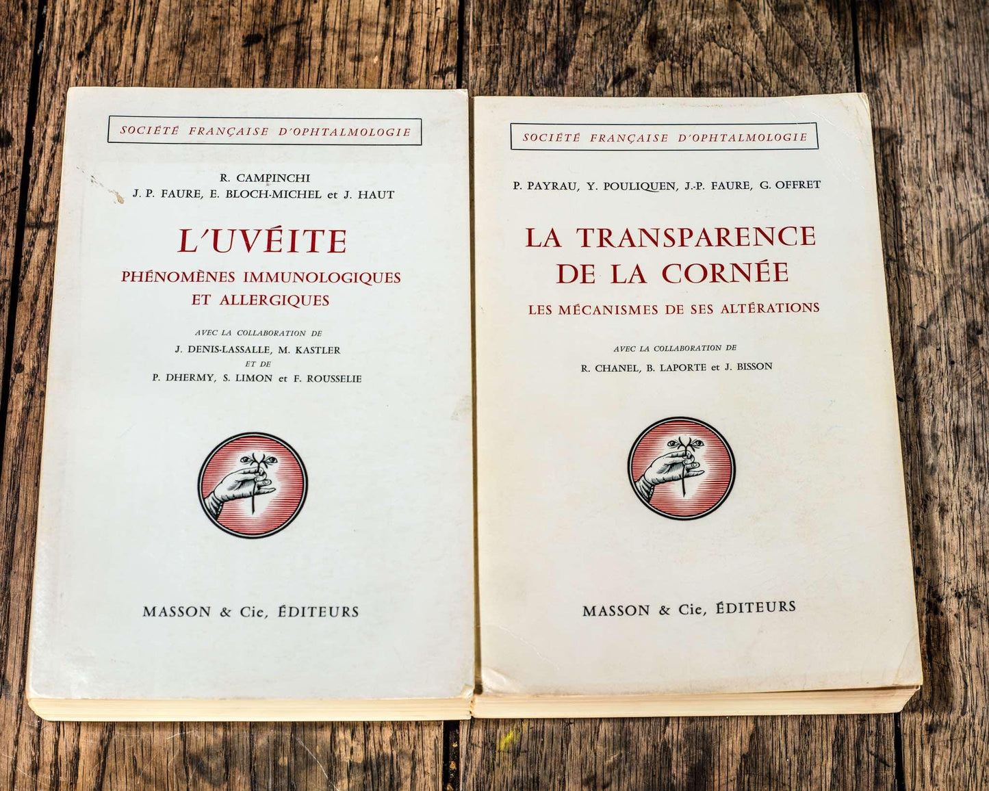
- UVEITIS, IMMUNOLOGICAL AND ALLERGIC PHENOMENA
AUTHORS:
R. CAMPINCHI, J.P. FAURE, E. BLOCH-MICHEL, J.HAUT, J. DENIS-LASSALLE, M. KASTLER, P. DHERMY, S. LIMON, F. ROUSSELIE
PARTS:
1. PATH IMMUNOPATHOLOGY. EXPERIMENTAL STUDY
2. CLINICAL AND ANATOMO-PATHOLOGICAL STUDY OF UVEITIS
3. ELEMENTS OF ETIOLOGICAL DIAGNOSIS
4. ETIOLOGY OF UVEITIS
5. THERAPEUTIC PROBLEMS
Report presented to the French Society of Ophthalmology on May 5, 1970
Masson & CIE, publishers 120, boulevard Saint-Germain , Paris (vi
PREFACE
We would first like to thank the Committee of the French Society of Ophthalmology and its Secretary General for entrusting us with the writing of this report.
As often happens to rapporteurs, we hardly anticipated, when this subject was proposed, all the difficulties we were going to encounter. This is to say all the gratitude we have towards those who helped us.
We thank First of all, our Master Professor GUY OFFRET: this work is his indirect work. He has, in fact, devoted a large part of his activities to the problems of uveitis, their immunological mechanism and their treatment. He knew how to interest us in it and guide us. Most of the guiding ideas in this report are those he has always taught. We have, moreover, benefited from all the facilities of his service, at the Ophthalmological Clinic of the Hôtel-Dieu de Paris , his files (more than a thousand cases of uveitis) and his laboratory. Let us express here all our gratitude and our respectful affection.
Our very warm thanks will also go to all the Ophthalmologists who
helped us :
Professor R. WITMER and Doctor ANNE-CATHERINE MARTENET (Zürich), Professor H. REMKY (Munich), welcomed us and advised us very friendly.
We owe many documents to Sir STEWART DUKE-ELDER, to Prof. E. S. PERKINS (London), to Prof. W. BÖKE (Kiel), to Prof. J. K. FRENKEL (Baltimore), to Prof. M. LANZIERI (Padua), to Prof. M. BLAGOJEVIC (Belgrade), to Prof. J. MICHIELS (Louvain), to Dr. I. M. DUGUID (London), to Prof. M. A. QUÉRÉ (Tours), to Dr. J. LAGRAULET (Paris). Dr. R. HEITZ (Haguenau) has very kindly entrusted us with his file on aqueous humor, which was very useful to us. Dr. H. A. MILLER and Mrs. Dr. M. A. LAROCHE (Paris) entrusted us with their observations of allergy to Candida albicans.
We would also like to warmly thank Professor R. WOLFROMM, the team at the Rothschild Hospital Allergology Center and particularly DTHs CL. VALLERY-RADOT and J. DENIS. The allergological methods that we use in the diagnosis and treatment of treatment of uveitis have been codified according to the principles developed by this School.
Father B. N. HALPERN, whom we thank warmly for his kind welcome, and his student Dr. MERI URGANCIOGLU, have shared with us their most recent work.
We We also owe a great deal of gratitude to Professor P. LÉPINE and the Virus Department of the Pasteur Institute, in particular to Mrs. Dr. J. VIRAT and Dr. J. MAURIN.
Mrs. Dr. G. DRACH, Research Officer at the C.N.R.S. , was kind enough to measure antistreptococcal antibodies for us.
Mr. G. SCHILLINGER helped us a lot with his knowledge of lens immunology.
Dr. A. FRIBOURG-BLANC advised us on the search for treponemas, Dr. F. MIKOL on neurosyphilis, Dr. A. PELTIER and the From B. AMOR on rheumatism, Dr. J. COUVREUR on toxoplasmosis.
Dr. G. DESMONTS and his collaborators (Toxoplasmosis Laboratory, Saint-Vincent-de-Paul Hospital, Paris) have shown us, for a long time - time, the interest of immunological research in toxoplasmosis; they have been providing us with valuable assistance for years in the diagnosis of this condition.
This report would not have been possible without the help of the Laboratory of the Ophthalmological Clinic of the Hotel-Dieu. We would like to thank in particular Professor Y. POULIQUEN, whose friendship was expressed once again on the occasion of this report, and Mrs. J. BISSON.
We owe a great deal to our devoted translators, Mr. J. CAMPINCHI, Dr. Y. LE TRAN and Mile Dr. Y. Z. KIM, as well as to Messrs. G. MARTINY and M. ESCOUBE who helped us to establish the bibliography.
We can never thank enough Messrs. CHIBRET, Dr. H. DECOUR and the staff of the Center of Documentation of the Chibret Institute, whose competence, welcome and friendliness have never wavered.
Our great gratitude goes especially to Mine MIROUFLE, librarian of the Institute of Ophthalmological Documentation of the French Society of Ophthalmology: sparing no effort or time, she made available to us the documentation that she manages with great skill. A work on ophthalmology with many references does not seem to us to be able to be carried out in Paris without her help. May she be particularly thanked.
We have found in Mr. MARQUISE a valuable and devoted collaborator. Several publishers have kindly authorized us to reproduce documents: let us especially mention The WILLIAMS & WILKINS Company, of Baltimore.
INTRODUCTION
T HE MAJOR DIFFICULTY of this report was due to the very imprecise limits of the subject: the title initially planned had been: "allergic uveitis". The term "allergic", too vague, can be taken in a restricted sense, essentially qualifying uveitis by immediate allergy or uveitis by microbial and tuberculin allergy; or, in a broader sense, also qualifying the classically autoimmune uveitis: sympathetic ophthalmia, phaco-antigenic uveitis; or finally, in a very broad sense, qualifying all uveitis, because a tissue hypersensitivity factor always seems to be more or less involved. This hypersensitivity factor is well demonstrated by experimentation. In clinical practice, it is difficult to detect. On the other hand, the detection of immune antibodies is often possible, for example in toxoplasmosis. Hypersensitivity and immunity are two responses, often inseparable, to antigenic aggression. We have therefore chosen the title: Uveitis; immunological and allergic phenomena.
It was not a question of describing all uveitis, especially on the clinical level, to redo what so many others have done well in the past and recently. We want above all to recall all the endocular immuno-pathological mechanisms that are found, variously grouped, in uveitis. We want to emphasize the clinical similarities that frequently result from this, and, thus, contribute to showing that few clinical and even anatomo-pathological aspects are clearly suggestive of a precise etiology; and that, practically, no clinical aspect is pathognomonic.
We will insist on experimentation, on immunological examinations, in particular on coupled examinations of aqueous humor and serum, to try to understand both the uniqueness and the diversity of uveitis. These examinations are, unfortunately, not yet numerous enough.
Experimentation, at first sight, seems very far from the clinic. What is there in common, in fact, between uveal irritation caused by injection of bovine serum albumin into the vitreous of the rabbit and clinical outbreaks of toxoplasmic or tuberculous uveitis? In fact, outbreaks of human uveitis have characteristics in common with secondary experimental uveitis (i.e. caused by a second or third injection of antigen in an animal sensitized by a first injection). Hypersensitivity undoubtedly plays a role in human clinical practice in most cases, and tion. Autoimmune factors are probably also involved, especially when the disease becomes chronic.
Clinical experience shows that the most common differences are in the uveitis: experimentation has focused on analyzing the share of each of these factors. However, it is difficult, based on chemical protocols, deliberately simplified to be demonstrative, to imagine a synthesis that fully explains human uveitis.
We must pay tribute to the German precursors of the early 19th century, who described the main types of uveitis caused by foreign proteins and who founded the autoimmune theory of certain uveitis, the eye being the first area of study for autoimmune phenomena. We must also pay tribute to Alan C. Woods, who devoted his life to uveitis, raising many problems in his long series of works: these problems have given rise to much research; this, without always confirming Woods' hypotheses, has made it possible to better delimit the questions. Finally, we must pay tribute to certain groups spread throughout the world, and in particular to modern American schools, thanks to which immunology, in some of its aspects, is today as advanced in ophthalmology as in other disciplines. experi-
The plan that we have followed includes:
1. The study of experimental uveitis and of endocular immunological mechanisms.
2. The clinical and anatomo-pathological examination of uveitis.
3. The etiological investigation of uveitis.
4. The etiological forms of uveitis. They have been chosen very arbitrarily from a certain number of causes that are best known in France, or in which immunological phenomena have been, to our knowledge, the most studied.
Thus, in the study of pyogenic uveitis, we have insisted on streptococcal allergy; among mycobacteria, we have studied tuberculosis and not leprosy; among protozoa, we have studied toxoplasmosis and not malaria, trypanosomiasis, amoebiasis, giardiasis; among helminthiases, uveitis by Toxocara canis and onchocerciasis, and not bilharzia, cysticercosis, ascariasis, filariasis; among mycoses, histoplasmosis and candidiasis and not other mycoses. Given the supposed analogies of their pathogenic mechanism, we have studied Reiter's syndrome, collagenoses and colopathies with rheumatism. For each etiology, we have made a final summary that would like to be practical.
5. Treatment. We emphasize drugs acting on inflammatory and immunopathological phenomena common to different uveitis.
Thus, we will speak very little about antibiotics, and will emphasize corticosteroids, ACTH, specific desensitization, the current possibilities of chemical immunosuppressants and anti-lymphocyte serum.
We have used quite a number of terms borrowed from immunological and allergological disciplines; these terms are not always familiar to ophthalmologists. Also, to facilitate reading, we have explained them in a glossary placed at the end of this work.
Finally, we will express a double wish:
First, that clinical ophthalmologists are not put off by the complexity of immunological phenomena that have been schematized as much as possible; we thus hope to make them aware of the amount of experimental work devoted to this subject.
On the other hand, to immunologists, particularly those who have studied ocular immunology, many of the concepts presented in this report may seem elementary. Others will seem superfluous, because they relate old and often outdated experiments. We believe that some of the old or incomplete works have their place in this work, the former because they testified to a new attitude at the time, the latter because it could be fruitful to take them up again. Our second wish will be that this report draws the attention of immunologists to the eye, a privileged organ of study. Our progress in the knowledge of uveitis currently depends largely on their assistance.
Number of pages: 972
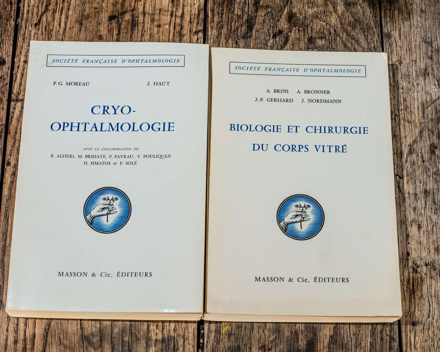
- CRYOPHTHALMOLOGY
AUTHORS:
P.G MOREAU, J. HAUT, R. ALFIERI, M. BRIHAYE, P. PAYRAU, Y. POULIQUEN, D. SIMATOS, P. SOLÈ
PARTS:
1. EFFECTS OF COLD ON TISSUES
2. CRYOSURGICAL EQUIPMENT
3. EXPERIMENTATION IN OPHTHALMOLOGY
4. OCULO-PALPEBRAL FROSTBITE
5. CLINICAL APPLICATIONS OF COLD
6. PRESERVATION OF OCULAR TISSUES BY COLD
Report presented to the French Society of Ophthalmology on May 4, 1971
Masson & CIE, publishers 120, boulevard Saint-Germain, Paris (vi
FOREWORD
THE IMPRESSION of having been able only imperfectly fulfilling, despite our efforts, the task entrusted by the French Society of Ophthalmology to present to its members a question as new as Cryo-ophthalmology, allowed us to measure the great honor that had been done to us. It is therefore with humility that we address to the Committee of the Society and to its Secretary General at the time, Professor DUBOIS-POULSEN, our very warm thanks.
Having been granted great freedom of maneuver, we broadened the subject and, from the Cryosurgery initially proposed, we slipped to Cryo-ophthalmology.
We also had total license in the choice of our collaborators, which allowed us to have recourse to the most eminent specialists in each field.
Me D. SIMATOS, Professor at the Institute of Applied Biology to Nutrition and Food (I.B.A.N.A.) in Dijon, is part of the small group of the world's great biophysicists of cold. We thank her for having been willing to make her knowledge clearly available to ophthalmologists.
Mrs. Doctor M. BRIHAYE-VAN GEERTRUYDEN, Professor at the Faculty of Brussels and scientific collaborator at the University of Amsterdam, has devoted important work to the action of cold on the sclera, the chorioretina and the vitreous. Her great knowledge of this subject led us to ask her to write this chapter. This was the occasion for fruitful discussions, and the birth of a friendship.
We thank Professor PAYRAU, for having written the chapter devoted to the preservation of ocular tissues by cold. This question is the subject of ongoing work carried out by the U86 Research Unit of I.N.S.E.R.M. (Ophthalmological Research Center of the A.-de-Rothschild Foundation, P' P. PAYRAU, and Laboratory of the Ophthalmological Clinic of the Hôtel-Dieu, Pr G. OFFRET) Destroying cells or preserving them by cold is the parent paradox of cryobiology, a true Janus, the double dispenser of life and death.
Professor Agrégé Y. POULIQUEN shared with us his knowledge and his work on the experimental action of cold on the cornea. Our friendships and our esteem will be enhanced by this collaboration. Let him be thanked here.
Professor Agrégé P. SOLÉ and Professor R. ALFIERI have carried out a long and particular experiment on the consequences of ocular cooling on the electroretinographic response; the lessons learned from it are rich. We thank them for their collaboration.
A large part of our recognition and our thanks goes to two of our masters:
To Professor G. OFFRET who, since 1964, has believed in cold in most of its applications; his communicative faith in Ophthalmology, his constant concern to improve surgical techniques, like the confidence he has constantly shown us, have been an irreplaceable support for us; a significant part of the iconography of this report comes from his collection.
To Professor L. PAUFIQUE, whose encouragement, critical spirit, surgical audacity and prudence have been invaluable to us.
This report would not have seen the light of day if Professor REY, a world authority on the physics and biology of cold, had not convinced us, a long time ago, of the interest of freezing applications. It was he who led us to work with the Société de l'Air Liquide, whose subsidiary, the Compagnie Française des Produits Oxygénés, we thank for its support during our past and current work. Since 1963, we have been able to study the problems of ophthalmic cryosurgery thanks to the infectious enthusiasm of an engineer from these Companies: passionate and gifted with great technical imagination, Mr. SCHULZ is thanked here for all the work he has accomplished for us, often during his leisure time.
Professor P. VINCENT, an eminent virologist from Lyon, very kindly gave us useful advice on corneal herpes. We thank him for this, as well as Doctor P. PICHON, assistant at the Dijon University Hospital, whose efficient and silent help was very useful to us.
We would like to express our gratitude to all the medical and nursing staff of the Ophthalmological Clinic of the Hôtel-Dieu in Paris, for the considerable clinical work carried out in this house for many years. Together with Doctor S. LIMON and Doctor J. A. BERNARD, they conducted an experiment on the chorio-retina, the conclusions of which are, in our eyes, fundamental.
Our gratitude goes to all our French and foreign colleagues who sent us the articles and documents in their possession concerning cryo-ophthalmology; we felt there how much ophthalmological friendship is a reality.
We express our deep gratitude to MM. F. THIERS, F. DREYFUS, M. LIMON, Mssrs. E. MOULIN and J. HAUT. They accepted the thankless task of translating the numerous German, Spanish and Italian publications.
The Documentation Center of the Chibret Institute has placed its traditional competence and helpfulness at our disposal to help us establish our bibliography and lend us rare works. We thank them for this, as well as, more particularly, Doctor H. DECOUR, Mile DELATTRE and Mrs. ERCOLANI.
The Institute of Ophthalmological Documentation of the French Society of Ophthalmology and its librarian, Ms. MIROUFLE, have contributed greatly to the writing of this report, by providing us with the works and photocopies that we needed. The dedication of Me MIROUFLE must once again be underlined and she should be thanked for it.
The help of Mr. MARQUISE has been indispensable and always effective for us
INTRODUCTION
In order not to break with tradition, the authors, by way of introduction, feel obliged to report on the difficulties they encountered in writing this work.
Indeed, when we were appointed as rapporteurs in 1966, the question of cold in Ophthalmology was presented as an avant-garde affair. However, requesting a report on a subject in the process of being cleared was contrary to the traditions of the French Society of Ophthalmology. The fact that it was a problem where the surgical part was essential contributed to breaking with customs and habits.
Designing the report according to classical standards was not possible. The novelty of the subject made inapplicable the usual technique of recalling old, classical and confirmed works, supported by modern observations and techniques.
Uncritical use of statistics would have been dishonest on our part: too often the enthusiasm of the first explorers of cryosurgery led them to set their operative successes at 100%. Such percentages, justifying the worst jokes about statistics, remove all educational value from them: they have become an end and not a means. These statistics can also be criticized for having been established globally for a specific condition, without going into the details of the clinical forms.
The opinions or comments noted in the literature should be corrected according to the instrumentation used. Indeed, too many poorly studied, and therefore dangerous, devices clutter the market. Incidents and operative complications will disappear when time has done its job of selection among these instruments.
Relying too much on experimental work was unrealistic given their rarity, even if in recent years an effort has been made in this direction. Outside of experimentation, cryosurgery was gradually, empirically, forging rules and methods. Cryobiology itself is still full of mysteries; its youth explains why our ignorance outweighs our certainties. In cryosurgery, application preceded knowledge, and one thinks of Picasso writing: I find first, then I search >.
New problem, new solutions.
Faced with the difficulties we have just listed, our conduct could only result in a commitment expressing our personal opinions. By setting aside all theory and all preconceived ideas, we have tried to be very pragmatic. The greatest prudence and the greatest scruples have guided us, because we know the moral responsibility we assume in exposing in the public arena new techniques held until now by a small number. On the other hand, the novice ophthalmologist in cryosurgery will be able to immediately grasp what is good and what is not so good in this new specialty.
The geographical distance has led us to a distribution of the writing, made according to our preferences or our previous orientations; this is why the name of the editor has been specified, especially since the necessarily subjective nature of the assessment of the action of cold has, for certain chapters of therapy, led to discussion and disagreement between the reviewers from different schools. It is rare that reviewers admit not to have always been in full communion of ophthalmological thought but it seemed to us more honest, and more profitable for all, to expose these bones of discord, in fact few in number. Is it useful to specify that this disagreement has not gone beyond the domain of certain surgical techniques? Undoubtedly, the Society's choice of authors was full of wisdom, because their many faults and their rare qualities clashed for the greater good of the common work.
Our ambition in this work was to go beyond the narrow framework of cryosurgery to address everything concerning the eye and cold.
In the first part we discover cold and cryobiology. The laws of cold will surprise some and will allow us to do justice to too many erroneous opinions currently accepted by ophthalmologists.
The cryogenic equipment used in our specialty is the subject of the second part. A specification of the ideal cryode is established, and many devices currently marketed are cited, with their advantages and disadvantages.
The experimentation constitutes the third part. Before describing it, structure by structure, a chapter is devoted to the penetration and diffusion of cold in the eye. The electroretinographic response to cooling is then reported.
Natural cold can, on the eye and its adnexa, cause damage whose severity is usually moderate. We have collected, we believe, all the known observations to synthesize the frozen eye. This clinical study forms the fourth part.
The fifth part is important because it deals with cold therapy. The stars are cryoextraction and retinal diseases, the supporting roles: iris hernia, spring conjunctivitis, corneal herpes. Vitreous cryosurgery is a surgery of the future. On the other hand, the treatment of hypertonia and tumors calls for many reservations.
The sixth part is reserved for a discipline of great future, the preservation of ocular tissues by cold.
We have eliminated from this report the endoocular metabolic modifications caused by generalized hypothermia, because they fall under a high physiological specialization; we have also eliminated pituitary cryodestruction for diabetic retinopathy, because its technical aspects are too far from our specialty.
The terminology, not yet codified and very much impregnated with English, shows the difficulty of our language in adapting to a modern scientific language.
seems more appropriate, but how can we use its adjective to say of a retina that it has been cryo-applied? We have therefore kept the improper terms which have the merit of already being consecrated by usage. Sometimes, usage will also go against syntax, because it is not possible to impose in all cases the hyphen after "cryo". Let us therefore admit that cryo-surgery and cryosurgery are written.
A happy recent habit is that a conclusion or summary ends each chapter. We have complied with it and, moreover, have translated it into English.
Cryo-ophthalmology is a new science, and this report is the first work in French devoted to it. This is why, whatever the value of the works cited, the bibliography has been as complete as possible. We stopped it in June 1970. It has been divided by part or chapter in order to facilitate the reader's research. The works concerning several chapters are obviously given as references after each of them. Many publications of a general nature concerning cold and the eye have been grouped together at the end of this introduction, with the list of films devoted to cryosurgery. We have, however, restricted as much as possible the references concerning known ophthalmological facts: this bibliography is above all that of cryo-ophthalmology.
Finding an apparatus, specifying a surgical tactic, comparing it to other methods, meeting other cold aficionados, was for the rapporteurs an exhilarating task, and the selfish opportunity to seal a friendship. We fear that the exposition of this cryosurgical passion has not been eloquent, but as the queen of a cold country, Christina of Sweden, said, only mediocre passions are eloquent >.
- BIOLOGY AND SURGERY OF THE VITREOUS BODY
AUTHORS:
A. BRINI, A. BRONNER, J.-P. GERHARD, J. NORDMANN
PARTS:
1. Biology of the vitreous body
2. Surgery of the vitreous body
Report presented to the French Society of Ophthalmology on May 14, 1968
Masson & CIE, publishers 120, boulevard Saint-Germain, Paris (vi
FOREWORD
The VITREOUS BODY is not, in general, the darling of the ophthalmologist, but the French Society of Ophthalmology apparently likes it very much. Has it not requested in a little over 30 years, 4 reports on this subject, those of KOBY, REDSLOB, BUSACCA, GOLDMANN and SCHIFF-WERTHEIMER and finally the one we have the honor of presenting today?
When, a few years ago, my friend Marc-Adrien DOLLFUS asked me to write the Report on the Biology and Surgery of the Vitreous Body, I hesitated a lot and for a long time. The subject is extremely vast and, despite the close relationships between the lens and the vitreous body, I had never specifically dealt with the latter.
However, I was able to realize very quickly that in few areas of our specialty such substantial progress had been made in recent times. They did not seem to be sufficiently known and above all there was a lack of an overall review that could constitute a starting point for new research.
However, the final acceptance was only possible after the agreement of my collaborators and friends BRINI, BRONNER and GERHARD. Their expertise naturally designated them for the histological and surgical parts of the report.
At no time have we forgotten the recommendations of the Secretaries General. We have therefore strictly observed the deadlines and abbreviated the text as much as possible, thus eliminating chapters that are of less interest to the clinician, for example, the chemistry of the embryonic period or others that are still little explored, which belong at least as much to retinal pathology - for example, the genesis of bridles. In the same vein, we have generally limited ourselves to very few species of mammals and have reduced the bibliographies whenever we could refer to good, easily accessible updates. Despite this, we fear that we have somewhat exceeded the regulatory number of pages.
We therefore plead guilty on this count, but we ask for extenuating circumstances, given that we had to deal with many problems far removed from the usual concerns of the ophthalmologist. Knowing his antipathy for formulas, we have deliberately renounced them; We have also spoken little about methods of investigation, but in return we have often had to go into detail and discuss at length. This explains why this report has become a little too long. We apologise for this, as well as for the omissions and gaps, which are inevitable due to the fact that we have been literally submerged by a flood of literature in recent years.
Two small innovations are intended to increase the "yield of the report":
1º At the top of each page of the text there will be an indication concerning the conclusions or the bibliography of the chapter. One will thus be able to very quickly realize the current state of the questions considered in the preceding pages and find all the documentation relating to them.
20 General conclusions recall some of the many questions of biology not yet resolved.
We hope that in this way this report will succeed in "hooking" the curious reader and that it will be useful to clinicians as well as researchers. It will thus have fulfilled the goal that each report pursues and which can be summarized in three words: Information - Orientation - Stimulation.
We wish to express all our gratitude to the Committee and the members of the French Society of Ophthalmology who have done us the honor of entrusting us with this Report. The subject was proposed to the votes of the General Assembly 5 years ago by the Secretary General at the time, Dr. Marc-Adrien DOLLFUS. Since that time, he has continued to be interested in it and to show us the most friendly goodwill. His successor, Dr. André DUBOIS-POULSEN, has greatly facilitated our task. We have found in him unfailing support and encouragement full of kindness in difficult times.
It is impossible for us to name all the colleagues who have lent us their assistance by sending us offprints or by their precise answers to our questions. We think of them with gratitude.
This feeling is particularly pronounced towards the great specialists, Professors E. A. BALAZS, of Boston and T. C. LAURENT, of Uppsala, who have allowed us to penetrate further into the vitreous body. We have never called upon their knowledge and experience in vain and we are keen to express here all the admiration we have for their talents as researchers and teachers.
Professor F. CLEMENT of Madrid has done us a great service by making available to us as soon as his report on the physiology and pathology of the vitreous body was published.
Professor A. PORTE of the Faculty of Sciences of Strasbourg has worked for many years in close and fruitful collaboration with one of us. This time again he participated very actively in histological research and was kind enough to introduce the presentation of the report with a general presentation on connective tissue. We have learned a lot from him and we hope that he will continue to give us his advice for a long time to come.
All the doctors of the clinic have helped us in various ways and Dr. R. HEITZ was kind enough to take charge of part of the chapter devoted to parasite surgery.
For their part, the Secretaries, photographers and designers did everything possible to enable us to finish within the time limits set for us. This dedication touched us greatly.
Strasbourg, October 1, 1967.
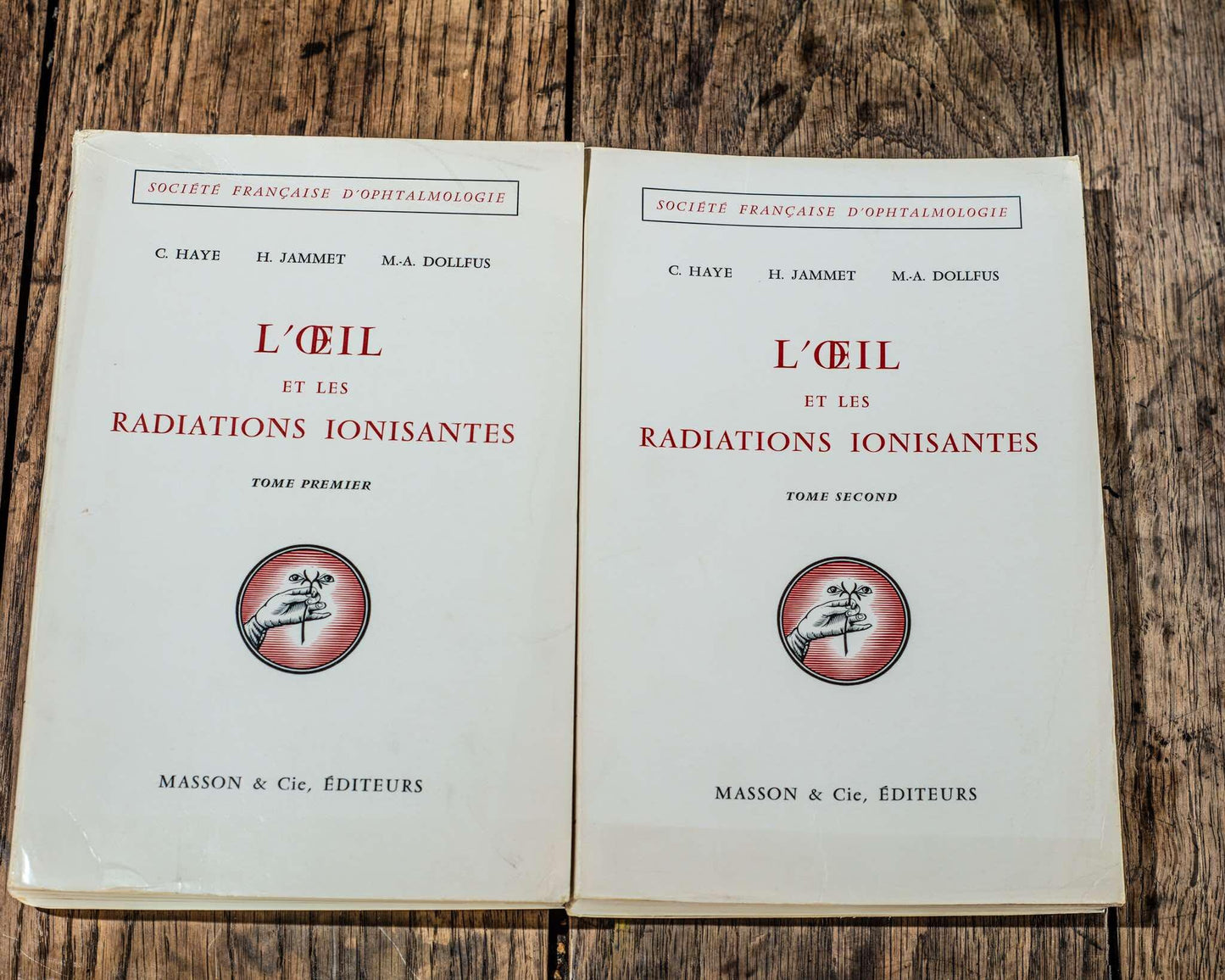
- THE EYE AND IONIZING RADIATION VOLUME ONE AND TWO
AUTHORS:
C. HAYE, H. JAMMET, M.-A. DOLLFUS
With 441 figures and 7 color plates
PARTS:
1. RADIOBIOLOGY AND RADIOPATHOLOGY
2. EYE LESIONS OF ATOMIC ORIGIN
3. ANALYTICAL STUDY OF THE EFFECTS OF DIFFERENT RADIATIONS
4. TREATMENT OF TUMORS
5. BETATHERAPY AND BUCKY RAYS
6. TREATMENT OF INFECTIONS AND DISEASES OF THE OCULAR APPARATUS
7. RADIOISOTOPES
8. FORENSIC MEDICINE AND CONCLUSIONS
Report presented to the French Society of Ophthalmology on May 11, 1965
Masson & CIE, publishers 120, boulevard Saint-Germain, Paris (vi
PREFACE
FOUR YEARS after the French Society of Ophthalmology has instructed my Master Félix TERRIEN to write a Report on X-rays and the eye, you were kind enough to ask me to deal with substantially the same subject; in the same session you granted me another perilous honor, that of succeeding my friend Guy OFFRET as General Secretary of our Society.
This was giving me at the same time a double and heavy burden that it was impossible to assume alone.
Since the time when Félix TERRIEN wrote his Report, the development of the use of ionizing radiation, the sudden onset of the atomic age, the better knowledge of radiation physics opened up new perspectives both for therapeutics and unfortunately also in the risks run by man, in particular in his ocular apparatus.
I therefore immediately looked among those around me for collaborators likely to carry out the work that we present to you today. From the outset, during the intermission following the business session, I asked my faithful student and friend Christian HAYE to kindly take on the writing of this Report. Without hesitation and perhaps without suspecting, at that moment, the enormous amount of work that he was going to take on with his usual dedication, he gave me his consent. Since that date, for five years, despite preparing for competitive examinations and multiple hospital assignments at the Cochin Hospital, the Curie Foundation and the Fontenay Research Laboratory, HAYE has provided an overwhelming amount of work. After gathering documentation that turned out to be more substantial than we thought, he set to work night and day on writing this work with, especially in the last year, the nagging concern of being ready on time. If you have this work in your hands, it is indeed to his perseverance and efforts that you owe it, and I express my deep gratitude to him. But if this subject had an ophthalmological basis, it was necessary, in order to apply the advice I have been giving for thirty years on the subject, to associate with our report a collaborator who is particularly knowledgeable about ionizing radiation. Some may have been surprised that I did not choose a pure radiotherapist, but that is because therapy only constitutes one part of our work.
I therefore asked my colleague and friend, from the Curie Foundation, Henri JAMMET, to kindly enlighten us with all his knowledge concerning the physics of radiation in general and more particularly the dangers they present for the eye, the means of protecting oneself from them, and also to explain to us what the sources of radiation are.
Henri JAMMET's dual membership, at the Curie Foundation as head of the Radioisotopes and Radiopathology department, and at the Atomic Energy Commission as head of the Health Protection Department, made him the person who seemed to me most suitable to help and advise us in this work, a whole part of which he wrote and the others of which he was kind enough to carefully reread. In this preface I would like to thank him very specially for having accepted this responsibility which he fulfilled with his smiling good grace and this despite absorbing occupations where his minutes are counted.
Faced with the work accomplished by these two friends, I had some qualms about adding my signature to this Report which cost me very little effort.
On the other hand, transformed into a two-faced Janus, Secretary General on one side, Rapporteur on the other, this split personality caused me some worries. During my four years as pro-consulate at the S.F.O. I have never ceased to preach the need for short reports not exceeding 600 pages and appearing before the Congress. As rapporteur I found myself faced with a subject which proved to be so vast and so important during the study that despite our efforts the recommended 600 pages have had little effect!!!. On the other hand, thanks to the efforts of my colleagues, you were able to read these two volumes before the Congress.
I therefore plead guilty on the first count but I believe that the present and future interest of the atomic era and the development of radiation treatments deserved not to hide any aspect of it from you. I hope that the work of HAYE and JAMMET will be well received by you and that you will forgive me for not having been too severe a censor.
Before closing this preface, I would be remiss if I did not recall that this work would perhaps not have been proposed to me if, one morning in 1933, Dr. PAULIN, then attached to the Curietherapy Department of the Curie Foundation, had not come to the Hôtel-Dieu to ask me to introduce him to ophthalmology and also to come and report to me at the Curie Foundation on the results of the treatments for eyelid epitheliomas. This is how our Monday afternoon ocular cancer consultation was gradually created, thanks to the support of the Directors of the Curie Foundation, Professor LACASSAGNE, and Drs. ROUX-BERGER and COURTIAL, and especially thanks to the precious and close collaboration of my old friend François BACLESSE and his assistants with whom I worked for more than twenty-five years.
Finally, I would like to express my deepest thanks to the directors of the Curie Foundation, to all my radiotherapist colleagues, to all the colleagues who have been kind enough to entrust their patients to us during this long period, allowing us to carry out this work which is thus somewhat theirs.
Marc-Adrien DOLLFUS.
INTRODUCTION
BEFORE PRESENTING THIS REPORT TO YOU, we would like to express our gratitude to the Committee and members of the French Society of Ophthalmology who have done us the honour of entrusting us with this work. If this subject was proposed to your votes five years ago, it was on the initiative of its dynamic Secretary General at the time, Professor Guy OFFRET. Since that date he has continued to be interested in this work, not only by opening wide the doors of his laboratory to us, but also by collaborating personally, despite his many occupations, in the experimental anatamo-pathological studies which without him could not have been done. We thank him sincerely for this.
In the minds of those who voted for this subject there was essentially the idea that it should be centered on ocular therapies by radiation. This question, certainly important, is infinitely too restrictive. With the development of the peaceful or military use of ionizing radiation, of increasingly precise biological and physical knowledge, it seemed necessary to us to consider broadly the whole of the question. After the chapters devoted to physical generalities, it seemed important to us to expose the ocular consequences of atomic explosions, the details of which had not yet been published in French. On this subject we believed it necessary to go beyond the question of ionizing radiation in a few pages, to describe the ocular complications from the same sources but caused by the other effects of the explosion. We would like to thank Mr. BYRNES in the United States and Mr. UTSUMI in Japan for the articles they sent us. In the following pages, we have studied the lesions caused by X-rays and gamma radiation on the various parts of the eye, then we have presented our personal experimental research before considering the therapeutic applications of ionizing radiation both in ocular cancerology and in certain inflammatory lesions. The use of radioisotopes in the diagnosis of neoplastic diseases of the globe was then described. Our work was already so voluminous that we were unable to include the use of radioisotopes in the physiological study of the eye.
If, as we said in the preface, one of us was the driving force behind these volumes, they nevertheless constitute a team effort, because all the chapters were reviewed by both authors, thus achieving full and complete collaboration.
The development of such a work could only be done with extensive documentation, gathered thanks to facilities and support from all sides and whose importance we would like to emphasize in this foreword. We express our gratitude to Mr. HAHN, Librarian of the Faculty of Medicine, to the Chibret Institute and to Mile DELATRE, as well as to Mrs. MIROUFFLE, of the Ophthalmological Documentation Center of the S.F.O. Thanks to them, we were able to gather a significant ophthalmological bibliography. We apologize if some omissions have been made. The work concerning atomic radiation and the study of ionizing radiation was provided to us by Mile VOLLOT and her Collaborators of the documentation group of the D.P.S. of the Nuclear Studies Center of Fontenay as well as by Mrs. ROULE, Attaché to the Atomic Research Center of Saclay.
The Atomic Energy Commission under the high direction of Mr. Professor Francis PERRIN, and more especially the Department of Health Protection headed by one of us, helped us considerably not only by providing us with this important documentation, but also and above all by opening an experimental research laboratory for us in the Nuclear Studies Center of Fontenay.
Our experiments were able to be carried out successfully thanks to the understanding of the administrative services of the Department and we particularly thank Messrs. BRESSON, ROUGÉ and Mrs. LEFRANC who smoothed out all the administrative problems that arose from the importance of a new laboratory.
This research required the collaboration of radiobiologists from the D.P.S. and we express all our gratitude to those who were interested in it: Professor AVARGUEZ, Commander LEGEAY, Captains COURT and PRAT. We also thank those who helped us in the technical realization of the irradiations: Messrs. DUPIN, DESAIVRE and GAUTARD. Mr. GAUDÉ was kind enough to establish the statistical study of our results. The anatomo-pathological sections and the microphotographs of the irradiated eyes were carried out at the Laboratory of the Ophthalmological Clinic of the Cochin Hospital then of the Hôtel-Dieu by the care of Mrs. BISSON and the diligent work of Mile MONTEIL, Mrs. CLÉMENT and Dr. PARTIOT. Our excellent friend and colleague Dr. POULIQUEN was kind enough to take charge of taking photographs of the corneal lesions under the electron microscope and writing the interpretation, and Dr. J.-P. FAURE was kind enough to review the biochemical chapter.
As part of this same experimental work, Doctor Colonel PAYRAU, Henri HAMARD and CUQ have kindly agreed to study the reactions of irradiated corneal grafts.
The chapters devoted to the therapy of ocular cancers by ionizing radiation are perhaps those that will attract your attention more specifically. Our experience on this point now extends over more than thirty years and this is not too long a period when it comes to cancerology. This experience was entirely acquired thanks to a final collaboration with all the departments of the Radium Institute (Curie Foundation) under the successive directions of Professor LACASSAGNE, Drs ROUX-BERGER and COURTIAL. At the Monday afternoon consultation, we first worked in contact with Mr. PAULIN and Mile BAUD during the initial period of curietherapy before the war of 1939, then for more than twenty-five years with our friends and colleagues Drs. BACLESSE, ENNUYER and ROUSSEAU and their assistants, the anatomo-pathological controls being assured in the department of Dr. GRICOUROFF. Dr. GONGORA was kind enough to take charge of the technical study concerning the Cobalt-60 disks. We also thank the Archives department of the Curie Foundation.
We owe a very special gratitude to all the colleagues who were kind enough to send us their work and their observations: MM. APPELMANS, F. BACLESSE, J. BARRAQUER, BLODI, BYRNES, CHARAMIS, DANA, ELLSWORTH, FOSSATI, J. FRANÇOIS, Mr and Mrs GUÉRIN, Mr HOLLWICH, Mrs LABORDE, Messrs LÉOZ, MANOLESCU, RAVERDINO, REEH, REESE, ROGUES, SABBADINI, STALLARD, STRAUB, UTSUMI and ZANEN.
But such a report must have a material structure and it is not easy to finish editing it within the time limit given by the S.F.O. to the Rapporteurs. Thanks to the work of our excellent secretary Mrs GRASSET, but especially to the considerable efforts of our editor, Messrs. MASSON and Co., and especially to the dedication of Mr. BAUCHER to whom we wish to express here our thanks and our gratitude, the edition of these two volumes was perfect materially and you were able to receive them on time.
In closing, we express to our Secretary General Dr. André DUBOIS-POULSEN all our thanks for his support and encouragement.
C. HAYE, H. JAMMET and M.-A. DOLLFUS. March 1, 1965.
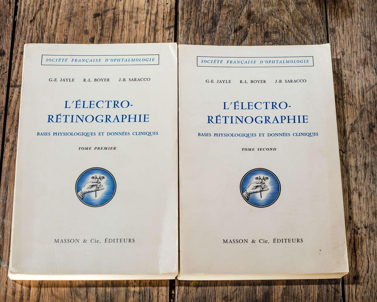
- ELECTRORETINOGRAPHY TOME PRIMIER AND SECOND
AUTHORS:
R.-L. BOYER, G.-E. JAYLE, J.-B. SARACCO
PARTIES:
PARTS:
1. GENERALITIES
2. ERG ACROSS THE ANIMAL SCALE
3. HUMAN PHYSIOLOGY
4. EQUIPMENT AND EXAMINATION TECHNIQUES IN CLINICAL ELECTRORETINOGRAPHY
5. SEMEIOLOGY OF DYNAMIC ELECTRORETINOGRAPHY
6. ELECTRORETINOGRAPHIC FUNCTIONS
7. ELECTRORETINOGRAPHIC DYSFUNCTIONS
8. ERG IN VARIOUS EYE DISEASES
9. ERG IN OPTICAL PATHWAY DISEASES
10. ADDITIONAL NOTIONS OF OCULAR ELECTROPHYSIOLOGY IN HUMANS
11. ADDITIONAL NOTIONS OF ELECTROPHYSIOLOGY OF THE OPTICAL PATHWAYS AND CORTEX OF MAN
12. THE PRACTITIONER FACED WITH THE VARIOUS MODES OF ELECTRICAL EXPLORATION OF THE OCULAR APPARATUS
Report presented to the French Society of Ophthalmology on May 12, 1964
Masson & CIE, publishers 120, boulevard Saint-Germain, Paris (vi
PREFACE
"Experience corrects man every day GETHE
THERE ARE MANY WAYS of approaching and presenting Electroretinography according to its clinical applications.
One consists of taking up all the ERG problems through an anarchic bibliography whose elements are presented as those of a mosaic of excavations incomplete: some facts are known, others are missing. The intermediate notions that would perhaps allow them to be linked together have not been defined.
This also involves penetrating the most diverse fields of animal and human physiology and pathology, passing through the most complex notions of nervous reactivity, physics and modern biochemistry that a mind, even a very educated one, can hardly embrace at the same time.
Despite these difficulties, MÜLLER-LIMMROTH has recently established a sum corresponding to this type of work and it therefore seems useless to follow a similar path.
Another approach, diametrically opposed, leads to considering clinical ERG from its purely practical aspect.
It leads to an impasse simply because no clinical interpretation is valid if one does not know the physiological characteristics of a normal response and its modifications when ocular parameters and stimulation parameters vary simultaneously.
We have chosen an intermediate path and written, not a compendium, but an essay designed from a certain number of precise general ideas.
These ideas have implied a choice and we have voluntarily sought to define the broad outlines of animal and human physiology, while removing a certain number of overly complex notions, in particular those concerning retinal purple.
The pharmacodynamic factors and data have only been touched upon. or micro-explo-
Conversely, a large place has been given to everything that concerns in normal man, the electrical exploration of the retina, the visual pathways and the occipital cortex.
*
From the physiological point of view, the problems posed by human physiology are, certainly, very particular, but cannot be separated from those posed by animal physiology. It was therefore natural to present in a way as schematic and as clear as possible the principal aspects of the ERG in animals.
For this purpose, the most characteristic ERG reactions of Invertebrates are studied, then selected, across the scale of Vertebrates, the most recent and most striking examples concerning the ERG of pure cone and pure rod cells whose study highlights the strong points but also the weak points of the classical theory of retinal duality.
In human physiology, we have tried to recall the classical notions, but also and above all, to gather the most recent data in order to show which path the study of pathological ERG can take. It is to physiology, in fact, and DANIS rightly emphasizes this in his monograph, that we must ask for openings on the future by borrowing its techniques while learning their limits.
From the technical point of view, all those of the authors who responded to the questionnaire that we sent to the different ERG laboratories were described in synoptic tables. It is regrettable, in this regard, that many of them did not respond or responded only very partially to our questionnaire.
Apart from these synoptic tables, the main types of techniques are presented in detail and more particularly the dynamic ERG technique that we have personally adopted.
Two very different levels are to be explored in Clinic.
First of all, the ocular level, which has already taken its place in current practice, and secondly, the level of the optical pathways and the cerebral cortex, which is still a level of research whose centerpiece is the EEG study of the responses of the occipital cortex.
It seems essential to us to identify, with regard to pathological ERG, a general notion on which we particularly want to insist with regard to the interpretation of the traces obtained.
Too many authors, it seems to us, think that these tracings can be interpreted through physiological concepts alone.
This is a point of view that we do not share. Physiology, as we have just said, has as its goal and normal limit to trace the frontier that separates the confines of the normal and the pathological.
If we refer to a Gaussian curve, these confines are located in the zone of sigma three, a zone in which physiological criteria can be applied to the assessment of pathological data.
We can admit, in fact, that, in this zone, pathology leads to modifications that only reflect the exaggeration or the reduction of the normal response.
It remains no less true that the pathological world is a world of dysfunctions and, incidentally at the terminal stage, of afunctions.
Dysfunction means distortion of physiological mechanisms, which, in the ERG domain, most often leads to the appearance of abnormal traces that no longer have, as a general rule, any characteristics of a physiological trace. This can result in the disappearance of any response.
It will therefore not be enough to measure such and such amplitude or such and such latency time of an abnormal trace to conclude that it is the modification of such and such wave or segment of a known physiological wave.
In fact, Pathology leads to the appearance of new electrogenetic phenomena, as soon as a function is very disturbed.
In the same way, a "spike-wave" complex in EEG has nothing to do with such and such cortical electrophysiological rhythm: it is
a new phenomenon due to the installation of particular disorders. Afunction means the absence of any electrogenetic response, either global or limited to certain parts of the trace.
The functions are of two types:
Congenital afunctions that modify only certain elements of the ERG response.
Acquired afunctions that are the ultimate stage of a dysfunction and that then result in electroretinographic silence.
The different parts of this book are presented in the classic mode:
Generalities, Animal Physiology, Human Physiology, Techniques, Human Pathology, Additional Notions of Ocular Electrophysiology. Additional Notions of Electroencephalography.
A final part is devoted to the problems that the birth and development in current practice of clinical ERG pose for the practicing physician.
Some parts have been more particularly entrusted to some of our Collaborators:
Animal Physiology to J. SARACCO; Techniques to A. REDON; Semiology to G. LE BRETON-OLIVEAU.
The complementary notions of ocular electrophysiology (electro-oculography and retinal electrovectorography) were developed by R. BOYER.
The study of the electrical responses of the optic pathways and the occipital cortex was entrusted to Dr. H. RÉGIS, Deputy Director of the Electroneurological Research Center of INSERM in Marseille.
Finally, Doctors M. ARNOUX, M.-C. BOFFA, J. FANTIN, Albert OURGAUD R. REYDY, M. RISS, and A. TASSY were kind enough to provide us with valuable and effective assistance in sorting and classifying the documents of the Marseille Ophthalmological Clinic.
R. BOYER took on the heavy responsibility for the bibliography and indexes.
In this field, the CHIBRET Documentation Center has provided us with a library, resources and technicians, including in particular, Mile DELATTRE, so active and efficient.
We are pleased to thank them in the most lively way for a collaboration without which our work could not have been carried out.
G.-E. JAYLE.
Number of pages: 1259
- THE NYSTAGMUS VOLUMES FIRST AND SECOND
AUTHORS:
M.-P. DELPLACE, J. HUDELO, P. LANTHONY, H. MARTERET, C. ROSSAZZA, Y.-M. RUELLAN, D. GODDÈ-JOLLY, A. LARMANDE, M. COLLARD, C. CONRAUX
PARTIES:
1.GENERAL
2.THE BORDERS OF NYSTAGMUS
3.SPONTANEOUS NYSTAGMUS
4.REVEALED NYSTAGMUS
5.INSTRUMENTAL NYSTAGMUS
Report presented to the French Society of Ophthalmology on May 8, 1973
Masson & CIE, publishers 120, boulevard Saint-Germain, Paris (vi
PREFACE
When the French Society of Ophthalmology did us the honor of entrusting us with the writing of a report on Nystagmus, it was with enthusiasm, and we realize this today Today, with temerity, we have accepted this mission. This problem seemed to us indeed very interesting and quite wrong known to most ophthalmologists.
But very quickly, we had to face the facts: this subject, which straddles disciplines as different as otology, neurology and ophthalmology, requires encyclopedic knowledge and we have were overwhelmed by the abundance and linguistic diversity of the bibliography, the reading of which alone would require an entire lifetime.
This is why it seemed reasonable to us to call upon the collaboration of informed specialists. We have therefore entrusted the writing of the chapters relating to neurological nystagmus and vestibular nystagmus to M. COLLARD and Cl. CONRAUX who, within the framework of the Strasbourg School, have devoted themselves for many years to this study at the Clinic Neurological (Pr ROHMER) and at the Oto-Rhino-Laryngological Clinic (Pr GREINER).
We did not hesitate, on the other hand, to call upon the assistance of our usual collaborators and of all our French and foreign Colleagues and Friends who were kind enough to share their experience with us, to communicate their publications and their observations or help us with translations.
In the field of bibliography, we would like to warmly thank the librarians of the Chibret Laboratories, the Ophthalmological Documentation Center of the Faculty of Paris and the Quinze-Vingts Library.
The goal that we The task we are assigned is relatively modest: we do not intend to write either a Treatise on Nystagmus or a Summary of Nystagmography. We have sought to define what a competent ophthalmologist must know, in our time, about nystagmus and of its multiple aspects.
Our report being produced under the aegis of the French Society of Ophthalmology, we thought it was necessary to deal in as much detail as possible with the three specifically ophthalmological nystagmuses: congenital nystagmus, optokinetic nystagmus and gaze nystagmus. eccentric. On the other hand, as regards otological and neurological nystagmus, we have asked our colleagues to aim rather at identifying the essential basic concepts, likely to be useful to us, as specialist practitioners. Although this report is a work team, we have made a point of indicating the names of the editors of each chapter in order to facilitate possible and enriching discussions.
We apologize for sometimes having had to go into somewhat tedious details and, to make up for it, We have decided to include at the end of the main chapters a summary of the points that seem to us to be the most important.
The imperatives of the edition forced us, at the last moment, to limit the importance of our already finished report, which obliged us, to our great regret, to delete the major part of our iconography, thus weighing down our text, to eliminate most of our observations and to pass quickly over the details of our personal experiences, referring the reader to the theses of our students.
D. GODDÉ-JOLLY and A. LARMANDE.
Number of pages: 1632
Language: french
